
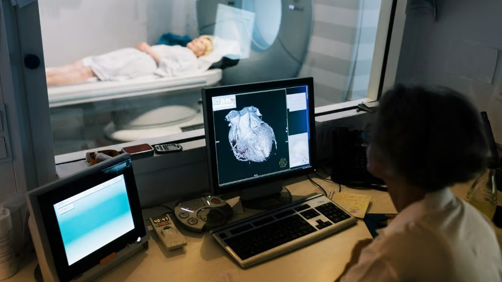
- A large study suggests that artificial intelligence (AI) measurements of heart function from MRI scans were slightly, but significantly, better at identifying those at increased risk of death than measurements made by experienced clinicians.
- The AI tool analyzed scans in approximately 2 to 3 seconds per patient, compared with around 13 minutes for expert clinical analysis.
- Although the AI tool demonstrated consistent performance, it had an approximately 1% error rate at the patient level, suggesting clinicians should continue to verify results and interpret findings.
- The findings support the potential use of AI as a first-line tool for cardiac MRI analysis, with expert review where necessary.
Cardiac MRI scans can provide detailed images of the heart, allowing clinicians to assess its structures and function. One key measurement is ejection fraction, which represents the proportion of blood pumped out of the heart’s main pumping chamber with each contraction.
Clinicians can calculate this by analyzing MRI images taken at different stages of the cardiac cycle to measure the volume of blood in the heart as it fills and contracts. This measurement can help assess heart function and guide decisions about treatment.
Now, research suggests that artificial intelligence (AI) may be more effective than clinicians at using heart MRI scans to predict patient survival, potentially helping speed up heart assessments and support clinical decision-making.
The study, published in
However, the researchers emphasize that clinicians should continue to review AI-generated results and interpret them in the context of each patient’s overall health.
AI measurements of heart function linked to survival
Researchers from City St George’s, University of London, and University College London (UCL) — both in the United Kingdom — analyzed MRI scans from 27,382 patients in the UK and the United States undergoing investigation for known or suspected heart conditions.
Participants were followed for an average of more than 5 years, during which 3,458 deaths were recorded.
The study focused on ejection fraction, which can help doctors determine whether a person may benefit from certain heart failure medications, implanted cardiac devices, or procedures to improve blood flow to the heart. It can also help assess surgical risk and monitor heart function during chemotherapy.
The team compared ejection fraction measurements produced by an AI tool with those made by experienced clinicians reviewing the same type of scans.
Their analysis found that AI-derived measurements were slightly but significantly better at identifying patients at increased risk of death. This association remained statistically significant after the researchers accounted for factors including age and sex.
“The improvement that we found was small, but across thousands of patients, small gains in precision could be meaningful, especially for patients at highest risk,” lead study author George Joy, MBBS, MRCP, an NIHR Clinical Lecturer at City St George’s, University of London, told Medical News Today. “Just as important, AI can perform in seconds, consistently, and at very low cost.”
“Ejection fraction is one of the most important measures of heart function,” Joy explained. “Doctors use it to decide on a wide range of treatments, for example heart failure medication and implantable devices.”
“We found that ejection fraction measured by AI was slightly more strongly linked to how long patients survive, than measurements made by doctors. AI measurements are fast and low-cost, and in some hospitals they’re ready before the patient has left the scanner. So AI could become the first-line measurement, with doctors reviewing it rather than manually it themselves, which is time consuming. That frees doctors up to focus on more complex tasks, such as interpreting the result alongside other test results and what they mean for the patient’s overall health.”
– George Joy, MBBS, MRCP
AI could analyze heart scans in seconds
In addition to its predictive performance, the AI tool was substantially faster than traditional clinical assessment.
The system could analyze a cardiac MRI scan in approximately 3 seconds per patient. Previous research by the same team found that expert clinicians typically required around 13 minutes to perform their analysis.
This difference could have practical implications for healthcare services, particularly as demand for diagnostic imaging increases.
If effectively implemented, AI-assisted analysis could help clinicians assess more scans in less time, potentially improving workflow and supporting more timely clinical decisions.
However, faster analysis does not necessarily mean that patients will receive treatment sooner or experience better health outcomes. Further research would be necessary to establish whether using the technology in routine clinical practice leads to measurable improvements in patient care.
Why clinicians still need to oversee AI results
Although the findings support the potential use of AI in cardiac imaging, the researchers stress that the technology should not replace clinical judgment.
The AI tool had an error rate of approximately 1% at the patient level. These findings suggest that the system performed consistently across the scans analyzed, but the researchers highlight the importance of checking its outputs.
“Errors were uncommon, affecting around 1% of patients,” Joy noted. “When they did happen, they led to ejection fraction (heart function) being either overestimated or underestimated. Both matter – that’s why a clinician should still review every result and remeasure where needed. We propose that AI does the routine measuring; the doctor checks it and interprets it.”
Cardiac MRI interpretation involves more than measuring ejection fraction alone. Clinicians must also consider other imaging findings, a patient’s symptoms, medical history, and results from additional tests when deciding on an appropriate course of treatment.
By automating a time-consuming measurement, AI could potentially reduce some of the workload associated with cardiac MRI analysis. It may also help standardize measurements between assessments.
Thus, the findings support an approach in which AI performs initial measurements, with clinicians reviewing results where necessary and retaining responsibility for clinical interpretation.
However, the study primarily examined the relationship between AI-derived measurements and subsequent mortality. It did not establish that introducing AI into clinical practice directly reduces deaths or improves treatment outcomes.
Further research will be important to determine how the technology performs across different patient populations, clinical settings, and MRI systems, as well as how best to integrate it into existing diagnostic pathways.
Combining AI with other heart tests
The research team plans to investigate whether combining AI-derived measurements from cardiac MRI scans with information from other tests could help identify heart problems earlier. One area of interest is the electrocardiogram (ECG).
Combining information from cardiac imaging and ECGs could potentially help identify abnormalities in the heart muscle before more substantial damage develops. Earlier identification may give clinicians an opportunity to investigate problems and consider treatment sooner.
Overall, the findings suggest that AI could become a useful tool for measuring heart function and identifying those at increased risk of death. However, further work is necessary to determine whether its use in routine care can translate into better outcomes for patients.