
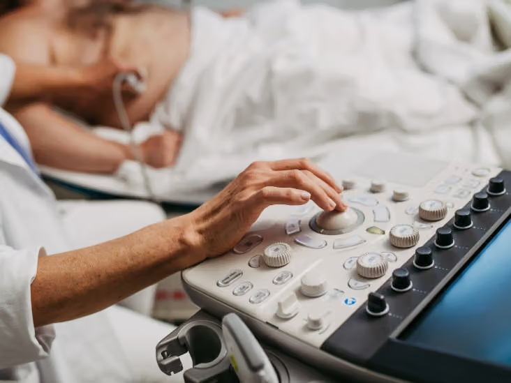
- A new AI model called DROID-RV can estimate right ventricular size to within about 3 millimeters and identify reduced function with high accuracy, potentially improving echocardiogram assessments.
- The AI model may help clinicians read echocardiograms faster, flag abnormal results, and identify patients who need additional MRI testing.
- Predictions from DROID-RV were associated with heart failure, atrial fibrillation, and mortality risk.
Artificial intelligence (AI) has the potential to improve the accuracy of echocardiograms of the heart’s right ventricle.
Researchers at the Mass General Brigham Heart and Vascular Institute and the Machine Learning for Health initiative at the Broad Institute of MIT developed DROID-RV, a deep learning AI tool, to test whether it could estimate the size of the right ventricle, as well as systolic function.
The study findings were published in October in
It was also able to identify reduced function of the right ventricle.
In a press release, lead study authors Mostafa Al-Alusi, MD, MS, and Patrick Ellinor, MD, PhD, explained the possible benefits of DROID-RV’s abilities in real-world clinical practice.
“Tools like DROID-RV can help echocardiographers read echocardiograms faster by providing preliminary measurements and flagging abnormal exams that need a closer look,” said Al-Alusi and Ellinor.
“They could also help identify patients who may benefit from additional testing with more expensive MRI scans to better characterize their RV, especially in situations where MRI machines or expert readers are scarce or in acute settings where immediate assessment is needed,” they added.
Highly accurate assessment of right ventricular function
The researchers described DROID-RV’s ability to identify the reduced function of the right ventricle with “high accuracy.”
The tool was also able to estimate the right ventricular diameter to within about 3 mm on average.
To begin testing the AI model, they trained it on more than 380,000 echocardiogram video clips from around 17,000 patients.
They then tested the model on 9,100 primary care patients within their own health system, as well as an independent dataset of 831 patients from a Hungarian hospital.
By comparing the model’s predictions with health records from the test data, they were able to determine how well the model could assess the function of the right ventricle, as well as whether predictions were linked to health outcomes later on.
Medical News Today spoke with Paul Drury, MD, board certified cardiologist, and associate medical director of electrophysiology at MemorialCare Saddleback Medical Center in Laguna Hills, CA, about the importance of assessing the right ventricle.
“Currently, cardiac MRI is the gold standard for RV evaluation; however, cardiac MRI is costly and not widely available. Echocardiography is inexpensive and widely available,” Drury said.
Drury also explained that the right ventricle is “very difficult to image on routine echocardiography. This is due to its complex, crescentic shape and anterior location in the chest, sitting immediately behind the sternum. Dilation of the right ventricle and right ventricular failure (decreased function) are associated with cardiac morbidity and mortality, so alternative methods to measure RV size and function are important.”
Drury also commented on the AI model’s ability to estimate ventricular diameter within around 3 mm.
“An accuracy of 3 mm is excellent for measuring cardiac chamber sizes, including the right ventricle, on an echocardiogram. This is acceptable for clinical practice,” he said.
Predictions associated with heart failure and atrial fibrillation
DROID-RV was able to predict whether larger right ventricular size or worse function of the right ventricle was associated with either heart failure or atrial fibrillation.
The AI model was also able to predict possible associations with mortality.
The study authors explain in the press release that predicted dilated right ventricles were twice as likely to result in mortality within 5 years compared with predicted normal right ventricle size.
Once they accounted for factors such as age, sex, and left ventricular function, the researchers continued to find an association between the model’s predictions and future cardiovascular disease.
“This is not surprising,” Drury said. “There are multiple causes of right ventricular enlargement and failure. Some of the most common
He added, “All of these chronic conditions, especially if left untreated, can lead to atrial fibrillation, heart failure, and subsequently death.”
Could AI benefit clinicians during echocardiograms?
While DROID-RV is currently not available as part of routine echocardiograms, Al-Alusi and Ellinor explained, “Eventually we may be able to use tools like DROID-RV to not only recreate echocardiogram measurements but also to better predict future cardiovascular disease.”
MNT asked Drury about the possible benefits of DROID-RV for clinicians if it were to become a routine part of echocardiograms.
“Due to the difficulty with accurately assessing right ventricular size and function with echocardiography, an AI model such as DROID-RV that would automatically calculate right ventricular parameters would alert cardiologists to potential issues with the right ventricle that would otherwise be missed,” explained Drury.
“This could lead to earlier diagnosis of the underlying condition (ie lung disease or sleep apnea), which could then be more expeditiously treated, or it could lead to additional targeted imaging with a cardiac MRI if right ventricular size and function needed to be confirmed.”
Drury also added that “I would need to see a study that confirms reproducibility of DROID-RV findings on echocardiograms done at a wide variety of cardiology sites with a wide variety of echocardiographers and wide variety of patients.”
He concluded, “Due to the variability of image quality of echocardiograms, I would need to see that DROID-RV could accurately assess RV size and function even on a poor-quality study. This would need to be compared to the gold standard, a cardiac MRI.”